|
Devices That Take Heart Pictures
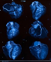
Equipment that is used to obtain heart pictures - cardiac imaging...is one of several methods used to help diagnose Coronary Heart Disease...The various pieces of equipment are collectively known as imaging tools or imaging modalities. Had a Cardiac Catheterization? It may be that certain symptoms are so obvious that test results are a mere formality...confirming and documenting the presence of the disease process. Sometimes symptoms may be a little more subtle...requiring some hard evidence one way or the other. In case you're wondering, the above heart pictures show several different angles of a 3D reconstruction of the surface of the heart. The white 'spiders legs' are the Coronary Arteries lying on the external surface of the heart muscle. Sometimes the diagnosis of Coronary Heart Disease is a chance finding...as a person might not have any symptoms at all...they are just presenting in the Doctors office for a routine medical, which may require a detailed cardiac assessment depending on who has requested the medical exam and the reasons for that. Of course, in all cases definitive diagnostic results assist in the recommendation of available treatment options...early detection can save lives as can early prevention strategies. Devices that obtain heart pictures...with the heart actually working in real time show... Different imaging technologies - devices used to take heart pictures - have their own strengths and weaknesses. Some technologies such as the Coronary Angiogram have been around for a long time...others such as the sixty-four slice Cardiac CT scanner are 'newer'...although are simply just an updated version of original CT scanner (old name CAT Scanner) technology that has been around since the 1970's.
The advantage of technologies that can obtain heart pictures is that they allow the specialist health care professional to evaluate hard evidence. Diagnostic tests such as the ECG and blood tests can point to cardiac issues, but there's nothing like having a picture right in front of you...where you can actually see structures working...or not working!
Coronary Angiography Probably the most well known test used to assess Coronary Heart Disease is the Coronary Angiogram. Coronary Angiography is a 'semi' surgical minimally invasive procedure used to access the coronary arteries and chambers of the heart using a coronary catheter - a meter plus long hollow tube that's only about two millimeters in diameter. Once access is gained to an artery in the leg or arm, the catheter is advanced internally along the chosen vessel to the target site. Have you been told you need to have a Cardiac Catheterization? Moving X-ray pictures are obtained and assessed for the presence of narrowed areas known as stenoses or a stenosis. This process can be used in both the diagnosis and treatment of Coronary Artery Disease.
read more about Coronary Angiography Click Here
Cardiac CTA You may have heard of Cardiac CT. The long name is Cardiac Computed Tomography Angiogram Scanner...quite a mouthful! This technology has been creating quite a buzz in recent times and has some real potential to displace the cardiac angiogram as the gold standard for heart artery and pumping chamber investigation. Further advancements are required in CTA though before coronary angiography no longer is the first choice of definitive investigation. The Cardiac CT scanner...which is actually a sophisticated X-Ray machine is able to obtain a huge number of heart pictures in multiple slices and recreate a three dimensional image using all the data it has collected. Medical staff can than view the heart on a monitor from any direction they like.... ...almost as good as holding the heart in your hands and tipping it from side to side to view all its surfaces. Cardiac CT scanners are also used to carry out calcium scoring procedures. In recent times this procedure has been marketed by Diagnostic Radiology and Cardiology clinics to symptom free members of the public as an 'anxiety reliever' if you are concerned about developing Coronary Artery Disease.
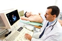
Cardiac Ultrasound Cardiac Ultrasound or Echocardiography is an ultrasound device that uses ultra high frequency sound waves to produce moving heart pictures of the heart chambers, valves, heart muscle and large blood vessels entering and leaving the heart. This device works in the same way as the ultrasound machine that is used for pregnancy scans. Cardiac ultrasound cannot be used to look at Coronary Arteries but it can be used to assess the damage caused to the heart muscle as a result of a heart attack. As far as a patient is concerned this examination is simple and painless...involving a device the size of a small microphone that is moved around on the surface of their chest to obtain optimal pictures of heart structures. Moving pictures can be recorded to hard disc or video and can be seen in real time on a monitor screen.
Stress Echo Stress echo is a cardiac ultrasound examination that records ultrasound pictures of the heart muscle, chambers and valves when the patient is at rest and directly after a period of controlled treadmill exercise to 'stress the heart'. Any difficulties that the heart muscle is having in coping with the demands of exercise are assessed.
IVUS Intravascular Ultrasound or IVUS is a different type of ultrasound examination altogether. IVUS is used in conjunction with Coronary Angiography to take ultrasound pictures of the inside walls of a selected coronary artery to gain cross sectional pictures of the artery in question. Especially useful in localizing and quantifying asymmetrical calcified plaque...inside a coronary artery that might not be shown accurately on a standard coronary angiogram.
Myocardial perfusion imaging Myocardial perfusion imaging is an examination that is performed in what is sometimes known as the nuclear medicine department of your local multi-disciplined hospital or clinic. This test procedure involves the detection in the patients blood of very low levels of an injected radioactive substance that is tagged to a pharmaceutical drug. Both these substances will then temporarily be absorbed by the heart muscle. The test is not invasive but does require a small injection to be given to the patient prior to or during the scan. Once the heart muscle has absorbed the radioactive material the patient is exercised on a treadmill...then lies down on an examination table where a large scanning device is place over their chest. The scanner detects faint radioactivity in the blood vessels of the heart muscle...the uptake of the injected substance by the heart muscle creates a pattern of distribution that is interpreted by the scanner and results in a picture that can tell medical staff how well the heart muscle is responding to its demand for oxygenated blood and nutrients.
Click here to go from Heart Pictures to Coronary Heart Health Home Page
Click here to go from Heart Pictures to Causes of Coronary Heart |


